
Our top priority is the care of your eyes. We want to keep your eyes healthy through regular eye health evaluations, communication, and education. This page lists a few of the most common eye diseases. Select from the following list of topics or scroll to learn about the causes, symptoms, and treatments for:
| Blepharitis | Cataracts |
| Conjunctivitis | Diabetic Retinopathy |
| Dry Eye Syndrome | Glaucoma |
| Macular Degeneration | Retinal Detachment |
Blepharitis
There are two types of blepharitis. Seborrheic blepharitis is often part of an overall skin condition called seborrhea, which may also affect the scalp, chest, back and the area behind the ears. The second form of blepharitis – staph blepharitis – is a more severe condition, caused by bacteria, that begins in childhood and may continue through adulthood.
Causes
Hormones, nutrition, general physical condition, and even stress may contribute to seborrheic blepharitis. Build-ups of naturally occurring bacteria contribute to staph blepharitis.
Symptoms
Blepharitis could be described as dandruff of the eyelids. Seborrheic blepharitis results in redness of the eyelids, flaking and scaling of eyelashes, and greasy, waxy scales caused by abnormal tear production. Staph blepharitis can cause small ulcers, loss of eyelashes, eyelid scarring, and even red eye.
Treatment
Careful cleaning of the eyelids can reduce seborrheic blepharitis. Application of hot packs to the eyes for 20 minutes a day can also help. Staph blepharitis may require antibiotic drops and ointments.

Cataracts
A cataract is a cloudiness that occurs in the lens of the eye. The lens is made mostly of water and protein that is arranged to let light through. Sometimes the protein clumps, blocking light and making the lens appear cloudy.
Symptoms
A person with cataracts may encounter faded colors, problems with light (such as halos, or headlights that seem too bright), poor night vision, double vision, or multiple vision.
Risk Factors include age, UV light, trauma to the eye, diabetes, smoking, excessive alcohol, steroid use, obesity and being a women.
Treatment
Your eye doctor can detect the presence of cataracts through a thorough eye exam, including a visual acuity test and dilation of the pupils. Cataracts are not completely preventable, but can be delayed, and at a certain point, surgery to replace the lens is routine.
Conjunctivitis
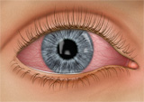 Conjunctivitis, commonly called pink eye, is a redness of the eye. It is often accompanied by a discharge (clear, yellow, or white) and itching in the eye.
Causes
Pink eye is most often a viral infection, but it may also be caused by bacteria or an allergic reaction. Viral pink eye is highly contagious.
Prevention and Treatment
To avoid spreading conjunctivitis, wash your hands often, do not touch the infected area with your hands, do not share wash cloths or towels, and avoid using makeup which may become contaminated. A child with pink eye should be kept from school for a few days. Sometimes an eye doctor will need to prescribe antibiotic eye drops and ointments to clear up conjunctivitis.
Diabetic Retinopathy
Diabetic retinopathy is a condition associated with diabetes. High levels of blood sugar may damage tiny blood vessels in your eye. New vessels may form to replace the damaged vessels. The new vessels can burst, resulting in blurred vision or even blindness.
Often no symptoms for diabetic retinopathy appear for a long time, however when they do they can include:
- "Floaters” – small specks that pass across your field of vision, made up of cells floating in the transparent gel of your eyeball
- Difficulty reading or seeing things close-up
- Sudden partial or total loss of vision or a shadow or veil in your field of vision
- Flashes
- Blurred or distorted vision
Risk Factors and Treatment
If you have diabetes, make sure you control your blood sugar level. This will reduce your risk of getting diabetic retinopathy. If you are experiencing some of the symptoms listed above, give us a call. If diagnosed properly, diabetic retinopathy can be treated with a laser procedure or a vitrectomy. Yearly dilated retinal exams are standard of care for the diabetic.
Dry Eye Syndrome
If your eyes are constantly gritty, dry or even watery you may have dry eye syndrome, which affects almost 10 million Americans. Dry eye syndrome is caused by a lack of, or poor quality of, tears. Tears lubricate the outer layer of the eye called the cornea. If the tears are not composed of a proper balance of mucous, water, and oil, the eye becomes irritated.
Symptoms
Dry eye syndrome leads to a number of symptoms, including grittiness, irritation, burning, excessive tearing, redness, blurred vision that improves with blinking, and discomfort after long periods of watching television, using a computer, or reading.
Risk Factors
There are many factors that can contribute to dry eye syndrome. These include dry, hot, or windy climates; high altitudes; air-conditioned rooms; and cigarette smoke. Contact lens wearers, people with abnormally dry skin, and the elderly are more likely to develop dry eye syndrome. You may also be more at risk if you take certain medications, have a thyroid condition, a vitamin-A deficiency, Parkinson’s or Sjorgen’s disease, or if you are a woman going through menopause.
Glaucoma
Glaucoma is a very common eye disorder affecting millions of Americans. It is caused by too much pressure on the inside of the eye. The fluid in your eyes helps to nourish and cleanse the inside of your eyes by constantly flowing in and out. When the fluid is prevented from flowing out, the intraocular pressure builds and damages the optic nerve. This causes a gradual loss in peripheral vision.
Symptoms
Those suffering from open-angle glaucoma experience a type of tunnel vision, where their field of vision gradually decreases. It can eventually lead to blindness. Narrow-angle glaucoma, which is rare, carries symptoms of sharp pain in the eyes, blurred vision, dilated pupils, and even nausea or vomiting. It can cause blindness in a matter of days, and it requires immediate medical attention.
Risk Factors
Heredity seems to be a risk factor. Also, you may be at greater risk if you are over 45 and of African descent or over 60 for the general population. You may also be at risk if you are diabetic, a high myope, have high pressures, have thin corneas or suspicious optical nerve appearance. Farsightedness is a risk for closed angle glaucoma. Finally, if you have used steroids or cortizone for a long period of time, or if you have suffered an eye injury in the past, you have a greater chance of developing glaucoma.
Macular Degeneration
Macular degeneration is a disease which affects a small area of the retina known as the macula. The macula is a specialized spot on the retina that allows us to see the fine detail of whatever is directly in front of us. Macular degeneration occurs when the macula begins to deteriorate.
“Wet” vs. “Dry”
Most often, macular degeneration is accompanied by formation of yellow deposits, called “drusen,” under the macula, which dry out or thin the macula. This is called “dry” macular degeneration. In rare cases, abnormal blood vessels develop under the macula and leak fluid. This is called “wet” macular degeneration.
Causes
A number of uncontrollable factors contribute to macular degeneration, including age, family history, sex (higher in females), race (highter in Caucasians), eye color, and farsightedness. Risk factors you can control include smoking, high blood pressure, exposure to harmful sunlight, obesity, inactivity and diet.
Symptoms
It is difficult for a patient to detect dry macular degeneration in its early stages. However, a retinal examination by an optometrist can find drusen, which are asymptomatic. The most common symptoms, when detected, include a spot of blurry vision, dark vision, or distorted vision. Wet macular degeneration progresses much faster than the dry variety. Both forms of macular degeneration can cause blindness.
Treatment
Currently, there is no cure for macular degeneration, but treatment is available to slow the effects. We often recommend certain vitamins that have shown to reduce the risk and help prevent the progression of macular degeneration.
Retinal Detachment
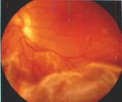 The part of the eye which collects light and transmits the light messages to the optic nerve and brain is the retina. It lines the inner back wall of the eye. When the retina separates from the back wall, it is known as retinal detachment. It is a serious condition which can cause permanent damage and vision loss if not treated quickly.
Symptoms
A retinal detachment often causes sudden defects in your vision. It may just cause a blind spot too small to notice, or it may cause a noticeable shadow which obscures your vision. An increase in “floaters,” which look like small particles or fine threads, may also be noticed. Finally, flashes of light are associated with retinal detachment.
Risk Factors
Eye injuries, tumors, and cataract surgery can cause retinal detachment. Near-sighted individuals and the elderly are at greater risk for spontaneous detachment. Also, diabetic retinopathy, a condition associated with diabetes, can cause bleeding which leads to retinal detachment.